
【Tech Support】﹤AI﹥Assisted Interpretation System for Chest Trauma Image
Kaohsiung Medical University / Prof. Liu, Hsin-Liang & Shih, Deng-Chiung
Pain Points Solved
Solves the challenges of interpreting chest X-rays where complex overlapping anatomical structures and subtle trauma lesions (such as fracture lines) lead to diagnostic oversight. It also addresses the high error rates and subjective variations in manual interpretation, particularly in emergency, primary care, or settings lacking specialized physicians.
Technology Introduction
The "Assisted Interpretation System for Chest Trauma Lesions" automatically identifies suspicious trauma-related lesions on chest X-ray images, providing visual annotations and confidence scores. The system comprises three core modules: a preprocessing module (using CLAHE and MSRCR to enhance contrast), an object detection module (utilizing YOLO), and a post-processing module that outputs visualized annotations and auto-generated summary reports.
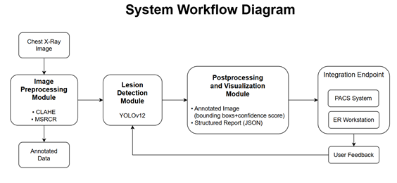
Figure 1. System Workflow Diagram
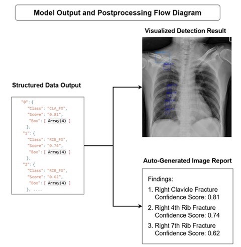
Figure 2. System Detection and Report Generation Diagram
Application Examples
- Emergency Medicine / Radiology: Real-time X-ray image interpretation for emergency trauma patients.
- Primary Care: Serves as a diagnostic assistance tool by highlighting suspicious lesion locations, reducing the risk of missed diagnoses due to a lack of human resources.
- Medical Education: A teaching system providing real-time comparison and feedback for residents and medical students interpreting X-rays.
- Telemedicine: Can be integrated with referral platforms for preliminary image interpretation.
Related Links
None
Industry-Academia / Tech Transfer Partner
None
Honors and Awards
None
Technical Contact
Mr. Hung, Assistant Manager
Kaohsiung Medical University
Tel: +886 7-3121101 ext. 2360
Email: R121084@kmu.edu.tw