【Tech Support】﹤AI、Smart Manufacturing﹥AI Algorithm for CT-to-3D Conversion
Kaohsiung Medical University / Prof. Ying-Hui Su
Pain Points Solved
CT images are often affected by metal artifacts, low contrast, and high noise, making it difficult to clearly identify anatomical structures such as bone, teeth, and root canals. This increases the complexity of diagnosis and treatment planning. In addition, traditional image segmentation methods largely rely on manual or semi-automated tools, which are time-consuming and highly dependent on operator experience, resulting in limited consistency and efficiency. Furthermore, existing workflows are typically confined to image processing or visualization stages and cannot be directly converted into clinically usable 3D models, thereby increasing the barriers for surgical guide fabrication and treatment planning.
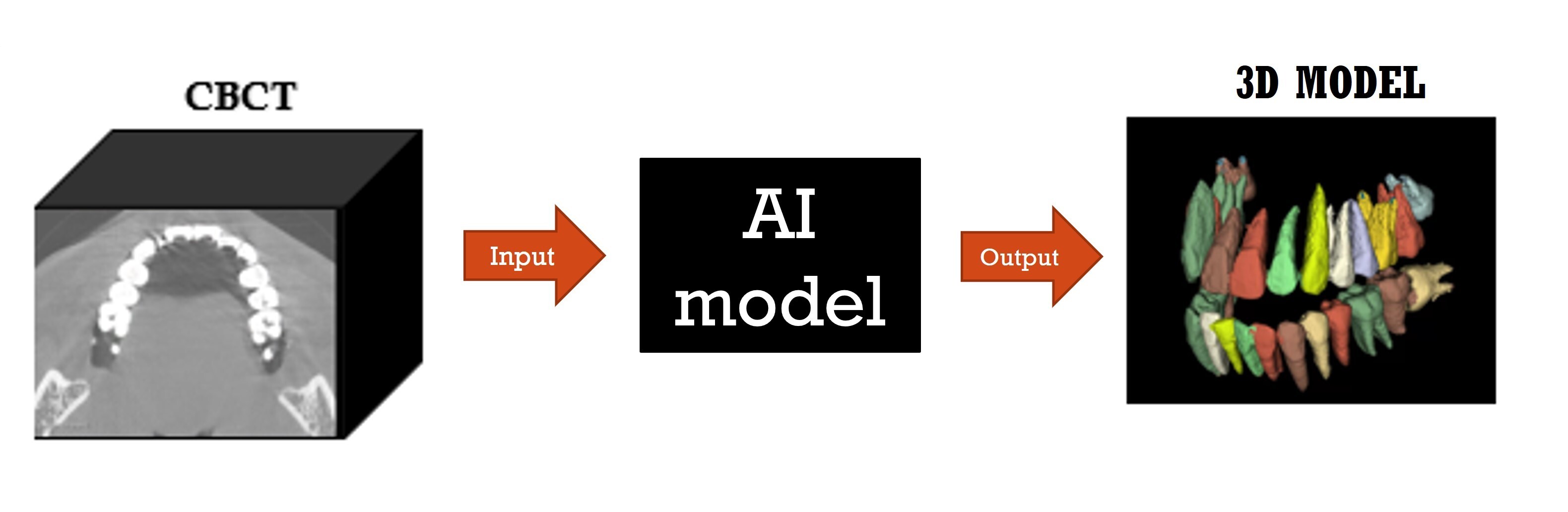
This technology utilizes a U-Net–based deep learning model to automatically segment bone, teeth, root canals, and artificial materials from CT images. It effectively reduces artifact interference and improves recognition accuracy, while enabling direct generation of 3D models for applications such as 3D printing and navigation-guided surgery. Overall, it establishes a fully automated workflow from imaging to clinical application, significantly enhancing efficiency and clinical feasibility.
Technology Introduction
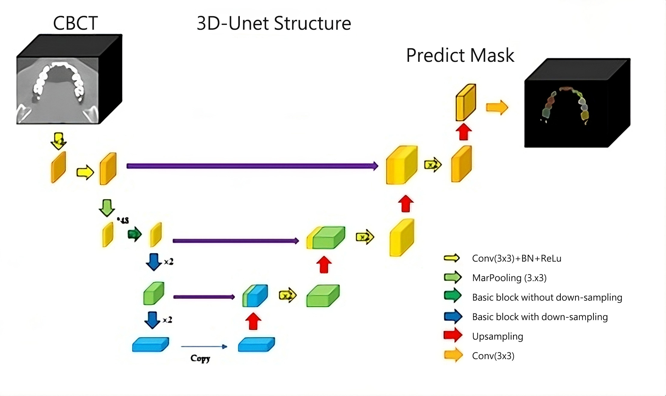
With the rapid advancement of artificial intelligence and deep learning technologies, convolutional neural networks (CNNs) have demonstrated great potential, particularly in medical image analysis. The U-Net model, a type of CNN, is widely adopted for medical image segmentation due to its powerful capability in feature extraction and reconstruction. It shows significant advantages in overcoming challenges such as metal artifacts and complex anatomical structures, greatly improving both accuracy and efficiency in image processing.
This patent describes an AI algorithm based on U-Net deep learning for automatic tissue segmentation in cone-beam computed tomography (CBCT). It enables the generation of accurate three-dimensional models by effectively segmenting bone, teeth, root canals, and prosthetic materials. The technology can be applied in various clinical fields, including orthodontic diagnosis, orthognathic surgery planning, 3D navigation-guided implant placement, guided endodontic treatment, and CT-guided autotransplantation of teeth.
Application Examples
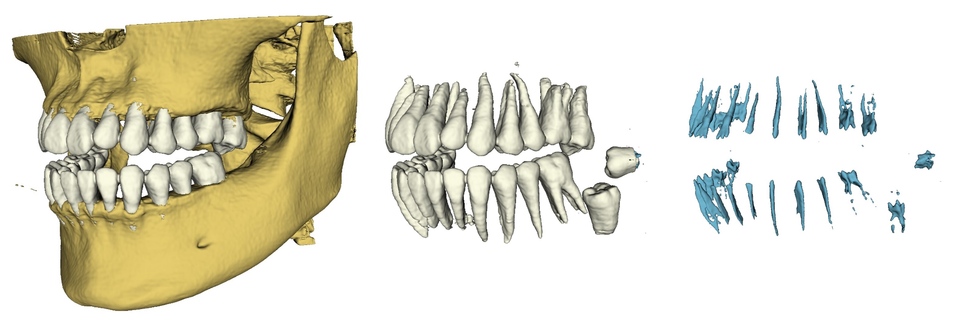
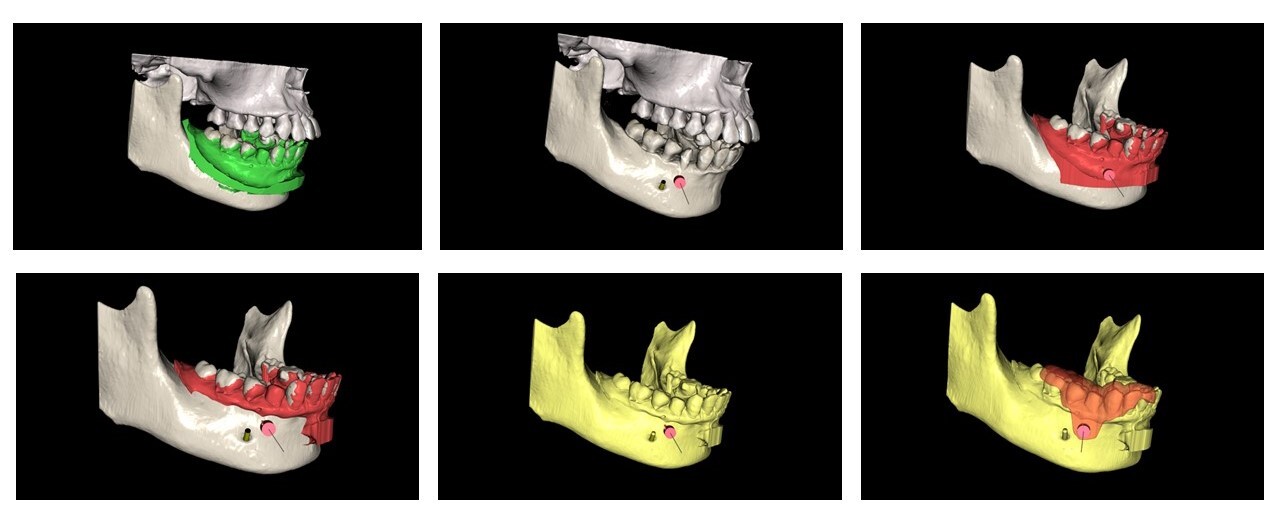
This case involves an apical surgery in which AI-assisted digital planning was implemented. First, CT images were converted into a three-dimensional model of the mandible. This model was then integrated with an intraoral scan of the dentition. The result is a fused model combining teeth from intraoral scanning and the jawbone from the CT surface. Based on this integrated model, a customized bone-supported surgical guide was designed and fabricated to guide the surgical position and direction.
Compared with conventional approaches, this workflow simplifies preoperative planning and improves localization accuracy. It also reduces the extent of bone removal and flap elevation during surgery, resulting in smaller wounds and shorter operative time, while enhancing overall treatment efficiency and stability.
Related Links
https://youtu.be/xvyR6emfAwc?si=o1eJJPWRflRvOxgd
Industry-Academia / Tech Transfer Partner
Industry-Academia Cooperation: muen AI
Honors and Awards
None
Technical Contact
Mr. Hung, Assistant Manager
Kaohsiung Medical University
Tel: +886 7-3121101 ext. 2360
Email: R121084@kmu.edu.tw